An Update on the CNS Manifestations of Neurofibromatosis Type 2
Coy S, Rashid R, Stemmer-Rachamimov A, and Santagata S.
Acta Neuropathol. 2019 Jun 4:1-23. PMID: 31161239
Available images
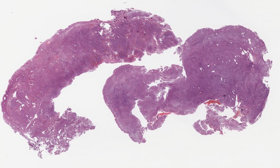
Vestibular Schwannoma
Vestibular Schwannoma
Image of Hematoxylin and Eosin (H&E) staining in a vestibular schwannoma tissue biopsy.
Image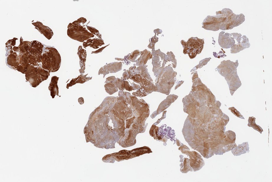
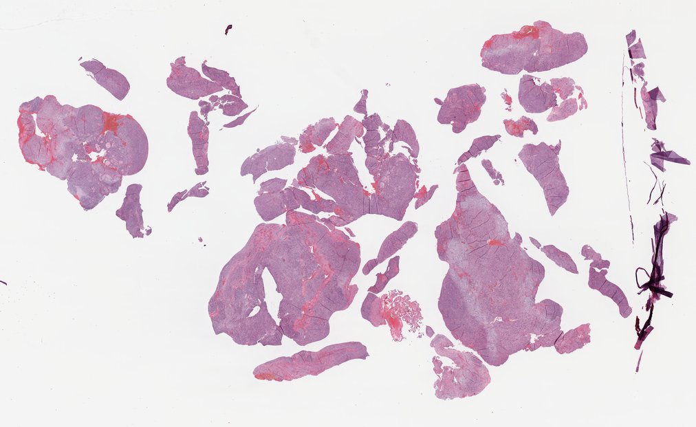
Vestibular Schwannoma
Image of immunohistochemistry (IHC) staining in a vestibular schwannoma tissue biopsy.
Image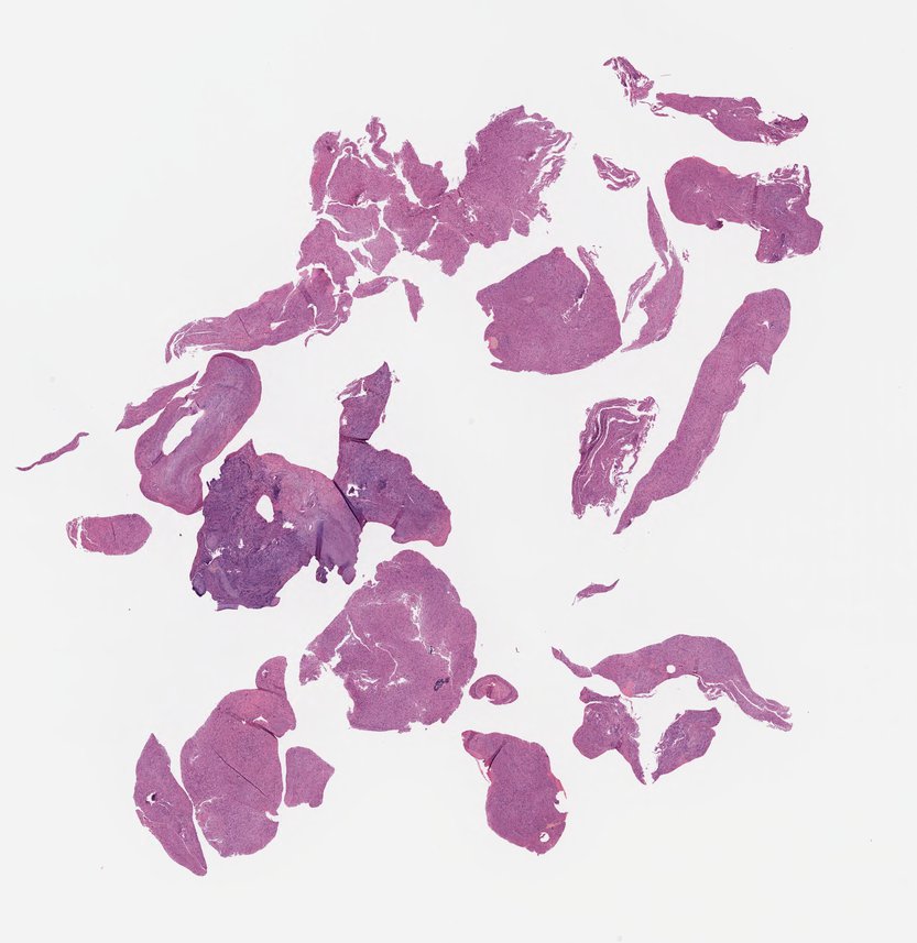
Vestibular Schwannoma
Image of Hematoxylin and Eosin (H&E) staining in a vestibular schwannoma tissue biopsy.
ImageNon-Vestibular Schwannoma
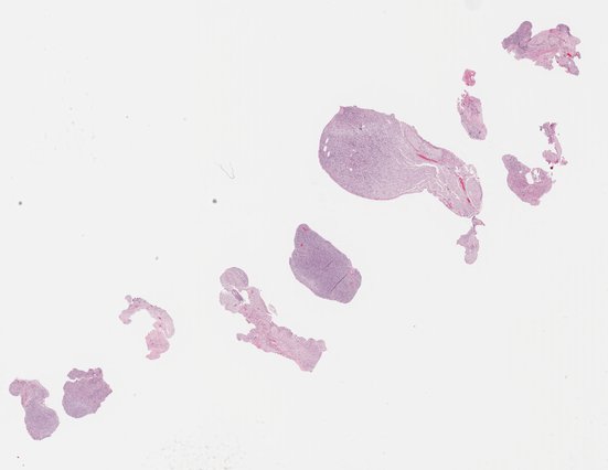
Schwannoma (spinal nerve root)
Image of Hematoxylin and Eosin (H&E) staining in a schwannoma tissue biopsy from spinal nerve root.
Image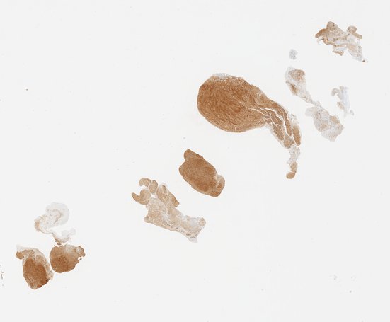
Schwannoma (spinal nerve root)
Image of immunohistochemistry (IHC) staining in a schwannoma tissue biopsy from the spinal nerve root.
Image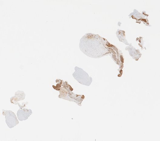
Schwannoma (spinal nerve root)
Image of immunohistochemistry (IHC) staining in a schwannoma tissue biopsy from the spinal nerve root.
Image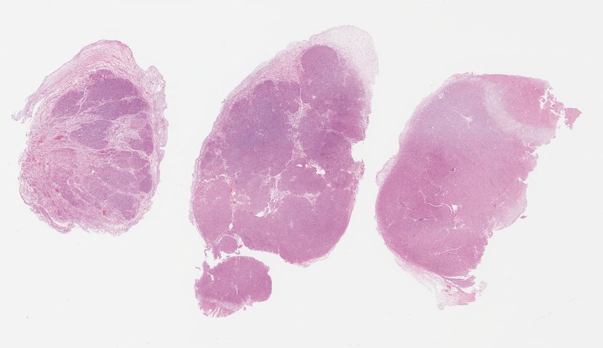
Plexiform schwannoma (larynx)
Image of Hematoxylin and Eosin (H&E) staining in a plexiform schwannoma tissue biopsy from the larynx.
Image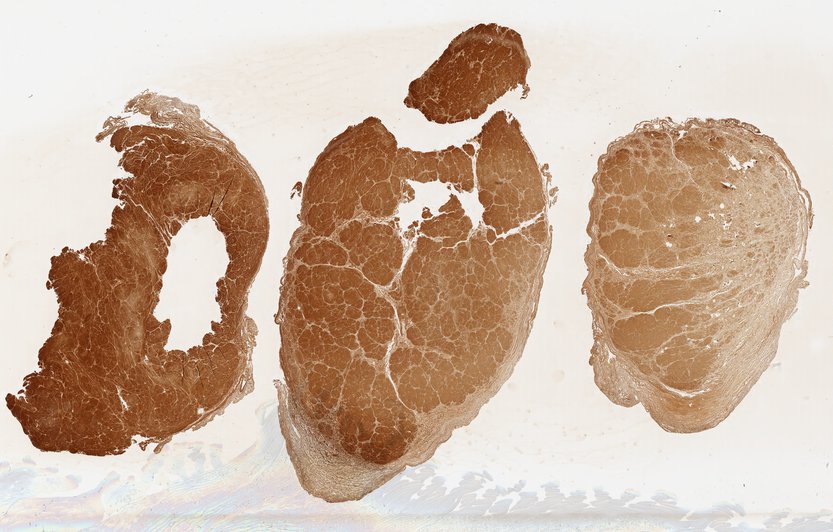
Plexiform schwannoma (larynx)
Image of immunohistochemistry (IHC) staining in a plexiform schwannoma tissue biopsy from the larynx.
Image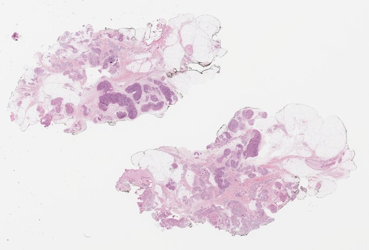
Plexiform schwannoma (finger)
Image of Hematoxylin and Eosin (H&E) staining in a plexiform schwannoma tissue biopsy from the finger.
Image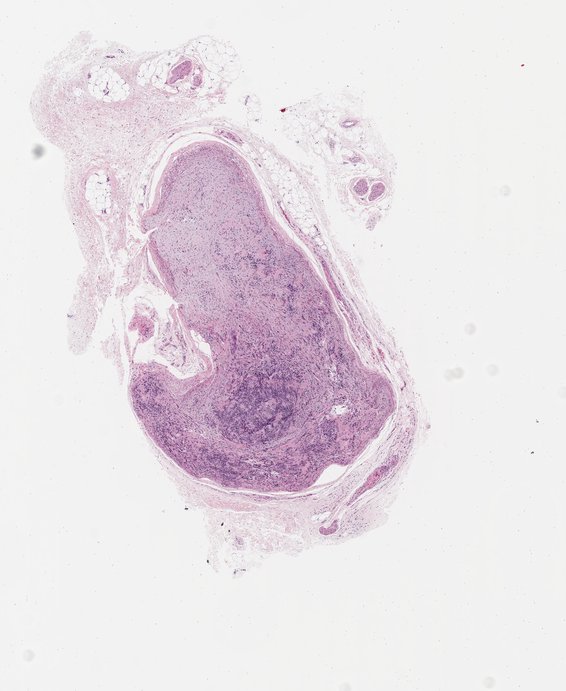
Intraneural schwannoma (finger)
Image of Hematoxylin and Eosin (H&E) staining in a intraneural schwannoma tissue biopsy from the finger.
Image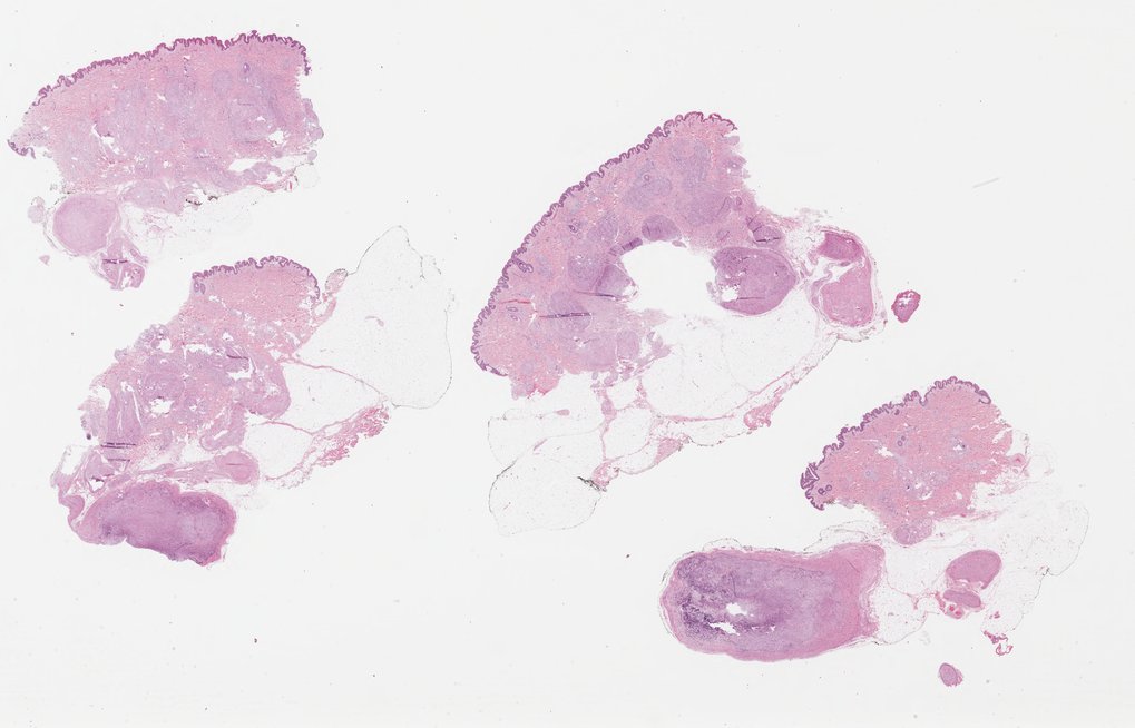
Plexiform schwannoma (soft tissue)
Image of Hematoxylin and Eosin (H&E) staining in a plexiform schwannoma tissue biopsy from soft tissue.
Image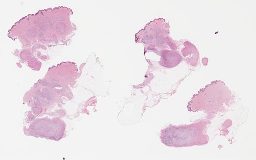
Plexiform schwannoma (soft tissue)
Image of Hematoxylin and Eosin (H&E) staining in a plexiform schwannoma tissue biopsy from soft tissue.
Image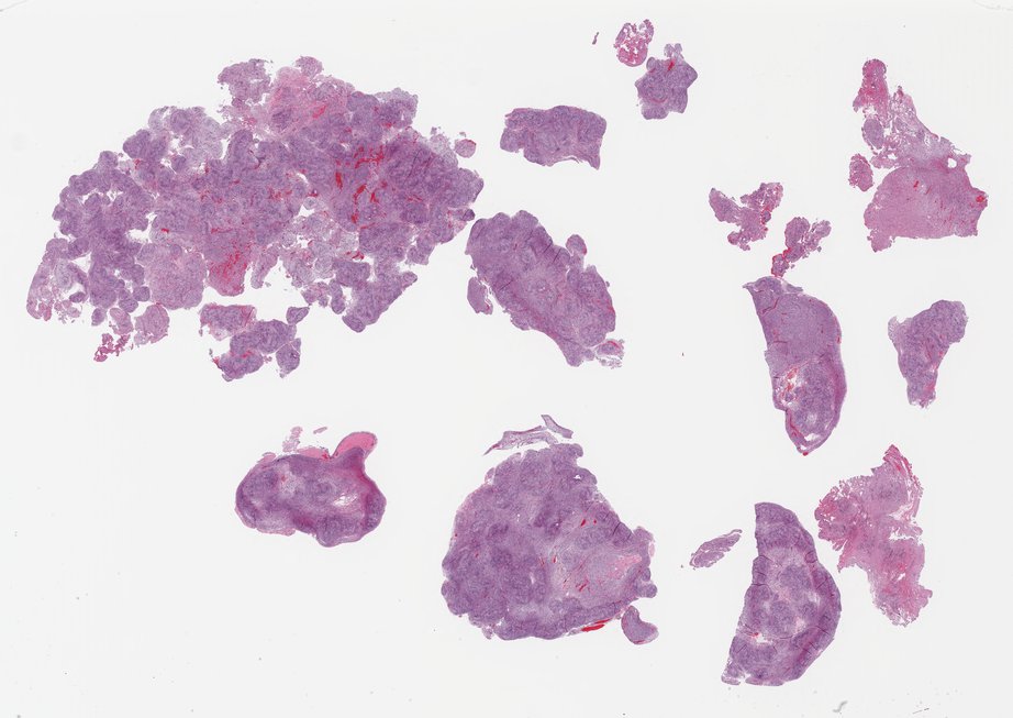
Plexiform schwannoma (spinal nerve root)
Image of Hematoxylin and Eosin (H&E) staining in a plexiform schwannoma tissue biopsy from the spinal nerve root.
ImageEpendymoma
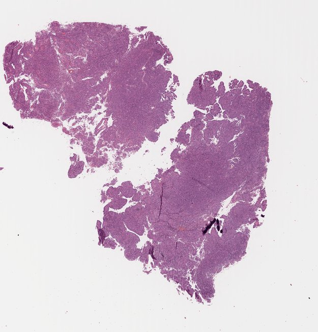
Ependymoma (4th Ventricle)
Image of Hematoxylin and Eosin (H&E) staining in an ependymoma tissue biopsy.
ImageMeningioma