Qualifying antibodies for image-based immune profiling and multiplexed tissue imaging
Du Z*, Lin JR*, Rashid R*, Maliga Z, Wang S, Aster J, Izar B, Sorger PK, Santagata S. (*co-1st author)
Nat Protoc. 2019 Oct; 14(10): 2900-2930. PMID: 31534232.
Raw Data | Publisher Page
Available images
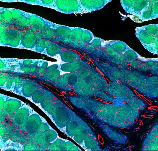
TONSIL
Representative t-CyCIF image acquired from a formalin-fixed, paraffin-embedded (FFPE) human tonsil tissue section stitched together using ASHLAR software from 224 fields acquired using a 40X/0.6NA objective.
CyCIF tonsil image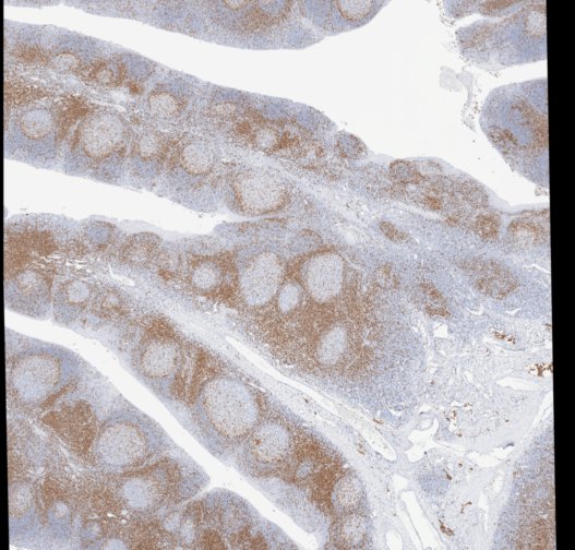
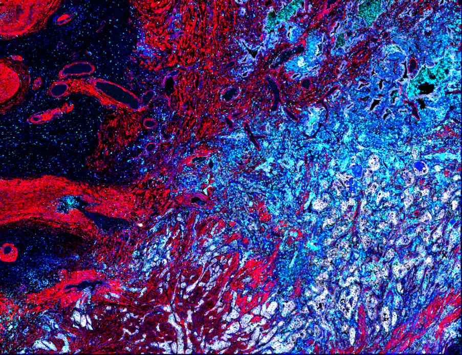
LUNG-3-PR: Primary lung squamous cell carcinoma
Representative t-CyCIF image of a squamous cell carcinoma of the lung stitched together using ASHLAR software from 132 fields using a 40X/0.6NA objective.
CyCIF lung-3 image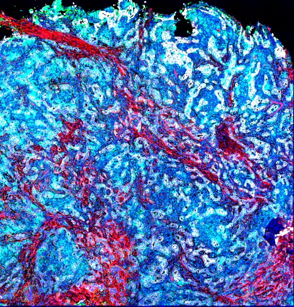
LUNG-1-LN: Lung adenocarcinoma metastasis to lymph node
Representative t-CyCIF image of a lung adenocarcinoma metastasis to a lymph node stitched together using ASHLAR software from 80 fields using a 40X/0.6NA objective.
CyCIF lung-1 image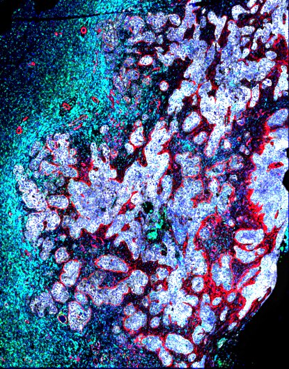
LUNG-2-BR: Lung squamous cell carcinoma metastasis to brain
Representative t-CyCIF image of a lung squamous cell carcinoma metastasis to the brain stitched together using ASHLAR software from 187 fields using a 40X/0.6NA objective.
CyCIF lung-2 image